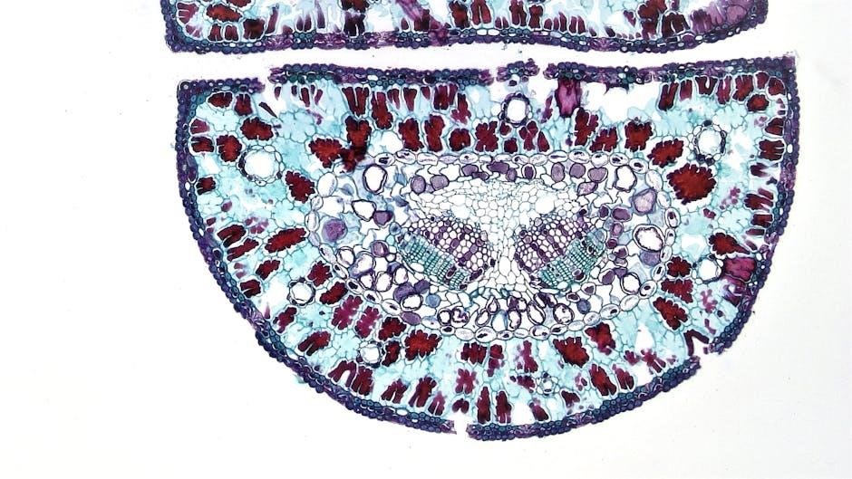
Plant cell coloring activities are engaging tools for visual learners, helping students remember cell structures and their functions through creative, hands-on practice․
1․1 Overview of Plant Cell Coloring Worksheets
Plant cell coloring worksheets provide a visually engaging way for students to learn about cellular structures․ These worksheets typically feature detailed diagrams of plant cells, labeling key organelles such as the cell wall, mitochondria, and chloroplasts․ By assigning specific colors to each structure, students can better differentiate and remember their functions․ Many worksheets include word banks and answer keys to guide learning and ensure accuracy․ They also often incorporate fun, interactive elements like matching games or short quizzes to reinforce knowledge retention․ These resources are ideal for biology classes, catering to visual learners and making complex concepts more accessible and enjoyable․
1․2 Importance of Coloring Activities in Biology Education
Coloring activities in biology education are invaluable for enhancing student engagement and understanding․ By transforming abstract concepts into visually organized diagrams, coloring helps students remember complex structures like plant cells․ It fosters active learning, making the study of cellular components more interactive and enjoyable․ Additionally, coloring exercises improve fine motor skills and hand-eye coordination while encouraging creativity․ These activities also serve as a practical assessment tool, allowing educators to evaluate students’ grasp of cellular anatomy․ Overall, coloring is a dynamic and effective method to simplify biology lessons, making them accessible and memorable for learners of all ages․
Materials Needed for Plant Cell Coloring
Printed plant cell coloring worksheets, colored pencils, markers, and a color guide are essential materials for this activity, ensuring students have everything needed to complete their task effectively․
2․1 List of Required Colors and Tools
For plant cell coloring, students need colored pencils or markers in specific hues like green for chloroplasts, orange for mitochondria, and grey for vacuoles․ Tools include sharpener, eraser, and a color guide․ A printed PDF worksheet is also necessary․ Ensure high-quality materials for vibrant results and clear labeling․ Proper tools enhance accuracy and creativity, making the learning experience enjoyable and effective․
2․2 Downloading the Plant Cell Coloring PDF
To begin, download the plant cell coloring PDF from reputable educational websites like biologycorner․com․ Ensure the PDF includes a diagram, color guide, and answer key․ Choose high-quality print settings for clarity․ Some worksheets offer interactive elements, while others are simple diagrams․ Verify the PDF includes structures like cell walls, chloroplasts, and vacuoles․ Print on standard paper for easy coloring; Accessing these resources online is straightforward, with many available for free․ Always check for an answer key to reference your work and ensure accuracy․

Key Structures of a Plant Cell
Plant cells consist of essential structures like the cell wall, cell membrane, cytoplasm, nucleus, mitochondria, chloroplasts, and vacuole, each serving unique functions․
3․1 Cell Wall
The cell wall is a rigid, outermost structure in plant cells, primarily composed of cellulose, providing support, protection, and maintaining cell shape․ It is semi-permeable, allowing certain substances to pass through while keeping others out․ Unlike animal cells, plant cells have a thicker cell wall, which is essential for withstanding osmotic pressure and supporting the plant’s structure․ In coloring activities, the cell wall is often highlighted in brown to differentiate it from the cell membrane․ This structure plays a critical role in plant growth and defense, making it a key focus in plant cell studies and educational coloring exercises․
3․2 Cell Membrane
The cell membrane, also known as the plasma membrane, is a thin, semi-permeable structure that encloses the cell and regulates the movement of materials in and out․ Composed primarily of phospholipids and proteins, it maintains cellular integrity while allowing selective transport of nutrients and waste․ In plant cell coloring activities, the cell membrane is typically shaded in orange to distinguish it from the cell wall․ This structure is vital for cell signaling and maintaining homeostasis, making it a fundamental component in plant cell diagrams and educational resources like the plant cell coloring PDF answer key․
3․3 Cytoplasm
The cytoplasm is the jelly-like substance within the cell membrane, hosting various metabolic processes essential for cell survival․ It consists of water, salts, sugars, and organelles like ribosomes․ Often colored yellow in plant cell diagrams, the cytoplasm provides a medium for chemical reactions and aids in transporting materials within the cell․ It plays a crucial role in maintaining cell shape and facilitating communication between organelles․ In educational resources like the plant cell coloring PDF answer key, the cytoplasm is distinguished by its light color, contrasting with the darker tones of organelles like mitochondria and chloroplasts․
3․4 Nucleus
The nucleus, often colored green in plant cell diagrams, is the control center of the cell, containing most of its genetic material in the form of DNA․ It is enclosed by the nuclear envelope, which is lined with nuclear pores allowing selective passage of materials․ The nucleus regulates gene expression, cell growth, and reproduction․ In educational tools like the plant cell coloring PDF answer key, the nucleus is prominently featured due to its importance in cellular functions․ Students typically color it distinctly to differentiate it from other organelles, ensuring clarity in understanding its critical role․
3․5 Mitochondria

Mitochondria, often colored orange in plant cell diagrams, are the powerhouses responsible for generating ATP through cellular respiration․ They are essential for energy production, converting glucose into usable energy․ In coloring activities, mitochondria are usually depicted as oval-shaped structures with inner folds called cristae․ The plant cell coloring PDF answer key highlights their importance, ensuring students recognize their role in sustaining cellular functions․ Coloring them distinctly helps students differentiate mitochondria from other organelles, reinforcing their understanding of cellular energy processes․
3․6 Chloroplasts
Chloroplasts, typically colored green in plant cell diagrams, are essential organelles responsible for photosynthesis․ Found in plant cells, they contain pigments like chlorophyll, which capture sunlight to produce ATP and glucose․ Their structure includes thylakoids, grana, and stroma․ In coloring activities, green shades highlight their role in energy production․ The plant cell coloring PDF answer key emphasizes their importance, ensuring students identify them accurately․ Chloroplasts are unique to plant cells, distinguishing them from animal cells, and are vital for food and oxygen production, making them a key focus in cellular biology education․
3․7 Vacuole
The vacuole, often colored grey in plant cell diagrams, is a large organelle responsible for storage․ It holds water, nutrients, and waste products, maintaining cell turgor and recycling cellular components․ In plant cells, vacuoles are prominent and play a key role in cell growth and maintenance․ The plant cell coloring PDF answer key identifies the vacuole as a storage compartment, ensuring students recognize its importance․ Its size and function distinguish it from other organelles, making it a critical part of cellular biology studies and a focus in plant cell coloring activities․
The Plant Cell Coloring Process
The plant cell coloring process involves matching colors to specific organelles, following a guide to ensure accuracy and enhance learning through visual engagement and creativity․
4․1 Step-by-Step Guide to Coloring
Begin by reviewing the plant cell diagram and color key․ Start with the cell wall, using brown, then the cell membrane in orange․ Next, color the cytoplasm light blue and the nucleus yellow․ For mitochondria, use orange, and chloroplasts green․ The vacuole should be grey․ Ensure each organelle is distinctly colored to avoid overlap․ Refer to the answer key for accuracy․ This methodical approach helps in understanding and retaining the structure and function of each cell part effectively․
4․2 Matching Colors to Cell Structures
Assign specific colors to each plant cell structure for clarity․ Use brown for the cell wall and orange for the cell membrane․ The cytoplasm should be light blue, while the nucleus is colored yellow․ Mitochondria are orange, and chloroplasts green․ The vacuole is typically grey․ Ensure each organelle’s color aligns with the provided key․ This consistent color scheme helps students differentiate structures visually and reinforces their functions․ Proper color matching enhances learning and ensures accuracy in identifying cell components, making the activity both educational and engaging․

Answer Key for Plant Cell Coloring
The answer key provides color and structure associations, ensuring accuracy․ It clarifies common mistakes, such as confusing cell wall and membrane colors, enhancing learning outcomes effectively․
5․1 Color and Structure Associations
The answer key outlines specific color assignments for each plant cell structure․ For example, the cell wall is typically brown, while the cell membrane is orange․ Mitochondria are often colored orange, and chloroplasts are green․ The nucleus is usually blue, with the nucleolus in a darker shade․ Vacuoles are commonly grey, and the cytoplasm is light blue․ These associations help students quickly identify and differentiate structures, reinforcing their understanding of plant cell anatomy through visual cues and consistent coloring standards․

5․2 Common Mistakes to Avoid
Students often confuse the cell wall and cell membrane, coloring them the same shade․ Ensure the cell wall is thicker and a different color, typically brown, while the membrane is orange․ Another mistake is neglecting to color the nucleolus darker than the nucleus․ Additionally, mitochondria should not be oversized, and chloroplasts must be distinctly green․ Vacuoles are often overlooked or incorrectly colored; they should be grey․ Properly following the color key prevents these errors, ensuring accurate representation and understanding of plant cell structures․ Attention to detail is crucial for both coloring and comprehension․

Beyond Coloring: Interactive Learning Activities
Engage students with labeling exercises, function matching games, and quizzes to reinforce plant cell knowledge, enhancing retention and understanding through interactive, hands-on learning experiences․
6․1 Labeling and Function Identification
Labeling and function identification activities complement coloring by enhancing understanding of plant cell structures․ Students match terms like mitochondria, chloroplasts, and vacuole to their respective roles, reinforcing knowledge retention․ These exercises often include short descriptions, encouraging learners to explain how each organelle contributes to cellular processes․ Interactive worksheets may also ask students to differentiate plant cells from animal cells, highlighting unique features like the cell wall and chloroplasts․ This hands-on approach helps students connect visual elements with functional concepts, making complex biology topics more accessible and engaging for visual and kinesthetic learners alike․
6․2 Comparing Plant and Animal Cells
Comparing plant and animal cells through coloring activities helps students identify key differences and similarities․ Plant cells feature a cell wall, chloroplasts, and a large vacuole, while animal cells lack these structures but may have centrioles․ Coloring exercises often include side-by-side diagrams, allowing students to visually distinguish these features․ Interactive guides may also highlight unique functions, such as photosynthesis in plant cells and mobility in animal cells․ This comparative approach enhances understanding of cellular diversity and reinforces recognition of organelles, making it easier for students to grasp complex biological concepts through visual and interactive learning methods․
Plant cell coloring activities effectively reinforce learning outcomes, providing visual mastery of cellular structures․ Additional resources, such as PDF guides and online worksheets, offer further exploration opportunities․
7․1 Summary of Learning Outcomes
Plant cell coloring activities enhance students’ understanding of cellular structures, aiding in visual and tactile learning․ By identifying and coloring organelles like mitochondria, chloroplasts, and vacuoles, students gain clarity on their roles․ This method improves retention, making complex biological concepts more accessible․ It also fosters a deeper appreciation for cell anatomy, encouraging curiosity and engagement․ The answer key provides a reference for accuracy, ensuring students grasp the correct associations between colors and structures․ This activity serves as a foundational tool for further exploration of cellular biology and related topics․

7․2 Recommended Worksheets and Guides
For effective learning, several plant cell coloring worksheets and guides are recommended․ The Plant Cell Coloring Worksheet from Biology Corner offers detailed diagrams and a comprehensive word bank․ Additionally, the Cell Coloring Guide PDF provides clear instructions and an answer key for accuracy․ These resources are ideal for students to practice labeling and coloring cell structures like chloroplasts, mitochondria, and the cell wall․ They also include interactive activities, such as comparing plant and animal cells, to enhance understanding․ These materials are available for free download and are suitable for various educational levels, ensuring a fun and educational experience․
