
Anatomy of Sports Injuries provides a detailed view of the body, aiding understanding of common athlete injuries and effective rehabilitation techniques.
This resource, featuring over 300 full-color illustrations, empowers fitness enthusiasts and athletes to navigate recovery and injury prevention.
It includes strength training exercises designed for common injuries, showcasing muscle impact and function, offering a pathway to self-guided physical therapy.
Understanding the Scope of Sports Injuries
Sports injuries are remarkably common, impacting individuals across all levels of athletic participation, from recreational fitness to elite competition. Understanding the breadth of these injuries requires acknowledging their diverse nature – encompassing acute traumas like fractures and ligament tears, to chronic overuse conditions such as tendonitis and stress fractures.
The scope extends beyond immediate pain; complications can arise if left unaddressed, hindering long-term athletic performance and overall quality of life. A comprehensive understanding, facilitated by resources like detailed anatomical guides, is crucial for both athletes and those involved in their rehabilitation.
These guides, often featuring full-color illustrations, illuminate the underlying anatomical structures affected, enabling informed decision-making regarding treatment and preventative measures. Recognizing the biomechanical factors contributing to injury, alongside appropriate rehabilitation exercises, is paramount for a successful return to activity and minimizing the risk of re-injury.
The Importance of Anatomical Knowledge for Rehab
Anatomical knowledge forms the bedrock of effective sports injury rehabilitation. Without a clear understanding of the involved structures – bones, muscles, ligaments, cartilage, and tendons – targeted and progressive treatment is impossible. Knowing precisely what is injured dictates the appropriate rehabilitation protocol, ensuring safe and efficient recovery.
Resources like detailed anatomical illustrations, found in guides dedicated to sports injury, are invaluable. They reveal how forces impact the body during exercise and pinpoint the specific tissues under stress. This insight allows for the design of strengthening and stretching exercises that address the root cause of the injury.
Furthermore, understanding anatomy empowers athletes to actively participate in their recovery, fostering a deeper connection to their body and promoting adherence to the rehabilitation plan. Ultimately, anatomical literacy transforms patients into informed partners in their healing journey.
Common Anatomical Structures Involved in Sports Injuries
Sports injuries frequently affect bones (fractures), muscles (strains/tears), ligaments (sprains), cartilage (meniscus damage), and soft tissues like bursae and tendons (bursitis/tendonitis).
Bone Fractures and Stress Fractures
Bone fractures represent a complete or incomplete break in the bone’s continuity, often resulting from acute, high-impact trauma during sports. These injuries require immediate medical attention, typically involving immobilization – casts or surgery – to facilitate proper healing and bone remodeling.
Stress fractures, conversely, are microscopic cracks developing over time due to repetitive stress, particularly common in weight-bearing bones like the calcaneus or tibia. Athletes engaging in high-impact, repetitive activities (jumping, running) are susceptible. Early recognition is crucial; continued activity can worsen the fracture.
Understanding bone anatomy – cortical and trabecular bone – is vital for comprehending fracture patterns and healing processes. Rehabilitation focuses on gradual loading, restoring bone density, and preventing re-injury through appropriate training modifications and biomechanical correction. Illustrated guides within resources like the Anatomy of Sports Injuries PDF aid in visualizing these concepts.
Muscle Injuries: Strains and Tears
Muscle strains and tears are common sports injuries resulting from overstretching or forceful contraction of muscle fibers. Strains involve damage to muscle fibers or tendons, categorized by grade (I, II, III) based on severity. Hamstrings, calf muscles, and biceps brachii are frequently affected.
Understanding muscle anatomy – fascicles, myofibrils, and the musculotendinous junction – is crucial for comprehending injury mechanisms. Eccentric muscle contractions, where the muscle lengthens while contracting, pose a significant risk.
Rehabilitation protocols, detailed in resources like the Anatomy of Sports Injuries PDF, emphasize the RICER protocol (Rest, Ice, Compression, Elevation) initially. Progressive loading with isometric and dynamic exercises restores strength and flexibility. Visual aids, including anatomical illustrations, demonstrate proper exercise technique and muscle activation patterns, aiding recovery and preventing recurrence.
Ligament Injuries: Sprains and Tears
Ligament sprains and tears occur when ligaments – the strong bands connecting bones – are stretched or ruptured due to trauma. These injuries are graded similarly to muscle strains (I, II, III) based on the extent of damage. Ankle sprains are particularly prevalent, often involving the anterior talofibular ligament (ATFL).
Anatomical knowledge of ligament structure and function is vital. Ligaments provide joint stability, and their injury often results from forces exceeding their tensile strength. Understanding biomechanical factors, like improper landing techniques, helps pinpoint injury causes.
Rehabilitation, as illustrated in resources like the Anatomy of Sports Injuries PDF, focuses on reducing inflammation with the RICER protocol and restoring joint stability. Immobilization may be necessary for severe tears. Progressive strengthening exercises, targeting surrounding muscles, are crucial for long-term support and preventing re-injury.
Cartilage Damage: Meniscus and Articular Cartilage
Cartilage injuries, encompassing both the meniscus in the knee and articular cartilage, present unique rehabilitation challenges. The meniscus acts as a shock absorber, while articular cartilage provides a smooth, low-friction surface for joint movement. Damage often occurs due to twisting motions or direct impact.
Understanding the limited healing capacity of cartilage is crucial. Unlike muscles and ligaments, cartilage has a poor blood supply, hindering self-repair. Anatomy of Sports Injuries PDFs emphasize early diagnosis and appropriate management.
Treatment ranges from conservative approaches – like strengthening surrounding muscles to improve joint stability – to surgical interventions like meniscus repair or cartilage grafting. Rehabilitation focuses on restoring range of motion, proprioception, and functional strength, often incorporating dynamic stretches and isometric exercises to protect the injured area.

Bursa and Tendon Injuries: Bursitis and Tendonitis
Bursitis and tendonitis are common overuse injuries affecting athletes. Bursae, fluid-filled sacs, cushion tendons and reduce friction around joints. Tendons connect muscles to bones. Inflammation of these structures – bursitis and tendonitis respectively – causes pain and limited movement.
Anatomy of Sports Injuries resources highlight how repetitive motions or sudden increases in training intensity contribute to these conditions. Common locations include the shoulder, elbow (tennis elbow), and hip. Accurate diagnosis, often aided by anatomical illustrations, is key.
Rehabilitation typically involves the RICER protocol (Rest, Ice, Compression, Elevation) alongside anti-inflammatory medication. Strengthening exercises, dynamic stretches, and gradual return-to-activity protocols are crucial to prevent recurrence. Understanding biomechanical factors and addressing muscle imbalances are also vital components of a successful recovery plan.
Specific Injury Locations & Anatomy
Detailed anatomical illustrations pinpoint injury sites – ankle, knee, shoulder, elbow, and hand – revealing crucial structures and biomechanics for targeted rehabilitation.
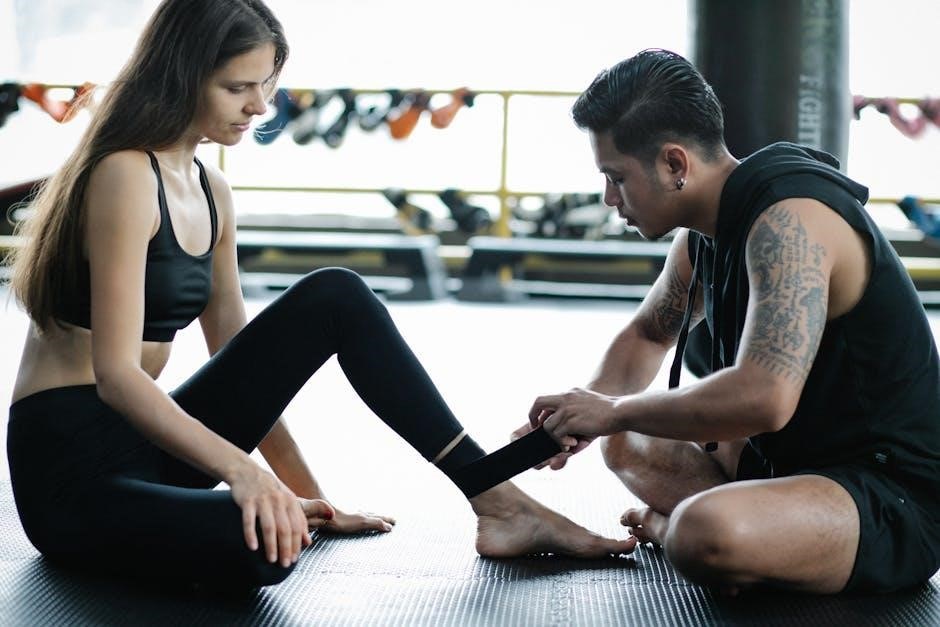
Ankle Injuries: Achilles Tendon & Calcaneus
The ankle joint, frequently injured in sports, centers around the Achilles tendon and calcaneus (heel bone). The Achilles tendon, connecting calf muscles to the calcaneus, is prone to tendonitis and rupture due to forceful plantarflexion or sudden stops.
Anatomy reveals the tendon’s limited blood supply, hindering healing. Calcaneus fractures often result from impact, like landing awkwardly from a jump. Understanding the surrounding ligaments – deltoid and lateral collateral – is crucial, as sprains commonly accompany these injuries.
Rehabilitation focuses on restoring range of motion, strengthening calf muscles, and gradually increasing load on the Achilles. Illustrations demonstrate proper stretching and strengthening exercises. Ignoring pain can lead to chronic issues, emphasizing the importance of early intervention and appropriate biomechanical assessment to prevent re-injury.
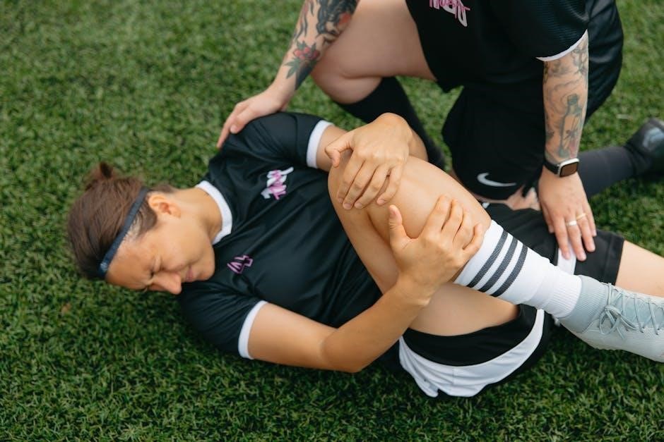
Knee Injuries: Femur, Fibula, and Joint Anatomy
The knee, a complex hinge joint, involves the femur (thigh bone), fibula (lower leg – stabilization), tibia (shin bone), and patella (kneecap). Injuries frequently target ligaments – ACL, PCL, MCL, LCL – crucial for stability. Meniscus tears, cartilage damage, are also common, often from twisting motions.
Understanding the femoral condyles and tibial plateau’s articulation is vital. The quadriceps and hamstring muscle groups provide movement and support. Illustrations detail ligament placement and function, aiding in diagnosis and rehabilitation planning.
Rehab emphasizes restoring range of motion, strengthening surrounding muscles, and proprioceptive training to regain balance and control. Ignoring knee pain can lead to chronic instability and osteoarthritis, highlighting the need for prompt, informed care and preventative exercises.
Shoulder Injuries: Humerus, Biceps Brachii, and Rotator Cuff
The shoulder’s ball-and-socket joint, formed by the humerus (upper arm bone) and scapula (shoulder blade), is prone to injury. The biceps brachii assists with elbow flexion and shoulder movement, but the rotator cuff – supraspinatus, infraspinatus, teres minor, and subscapularis – provides crucial stability and rotation.
Common injuries include rotator cuff tears, impingement syndrome, and shoulder dislocations. Detailed anatomical illustrations showcase muscle attachments and joint mechanics, essential for understanding injury patterns.
Rehabilitation focuses on restoring range of motion, strengthening the rotator cuff muscles, and improving scapular control. Progressive exercises, starting with isometric contractions and progressing to dynamic movements, are key to a successful recovery and preventing future instability.
Elbow Injuries: Forearm and Extensor/Flexor Muscles
The elbow joint relies heavily on the coordinated function of forearm muscles – extensors on the lateral side and flexors on the medial side. These muscles attach via tendons to bony prominences, making them vulnerable to strains, tendinitis (tennis or golfer’s elbow), and even tears.
Understanding the anatomy, including the radial and ulnar collateral ligaments, is crucial for diagnosing and treating elbow pain. Detailed illustrations highlight muscle origins, insertions, and actions, aiding in targeted rehabilitation.
Rehabilitation protocols emphasize restoring pain-free range of motion, strengthening forearm muscles, and addressing any biomechanical imbalances. Eccentric exercises are particularly effective for treating tendinopathies, promoting tendon healing and preventing recurrence.
Hand and Finger Injuries: Joint Dislocation & Muscle Damage
The hand and fingers, complex structures with numerous small joints and intrinsic muscles, are prone to injuries during sports. Dislocations, often occurring from direct impact or forceful twisting, disrupt joint alignment and require prompt reduction.
Muscle damage, including strains and contusions to the thenar and hypothenar eminences, can impair grip strength and dexterity. Understanding the anatomical arrangement of flexors, extensors, and intrinsic hand muscles is vital for accurate diagnosis.
Rehabilitation focuses on restoring joint stability through strengthening exercises, regaining range of motion with targeted stretches, and protecting healing tissues; Splinting or taping may be necessary to support injured joints and prevent re-injury during activity.
Injury Mechanisms & Biomechanics
Understanding forces, impact, and eccentric muscle contractions is crucial for pinpointing injury causes. Biomechanical factors significantly contribute to sports-related musculoskeletal trauma.
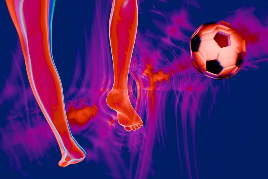
Force and Impact on the Body
Sports injuries frequently arise from the body’s response to external forces and impacts. These forces, whether direct (like a collision) or indirect (resulting from rapid deceleration or acceleration), can exceed the structural capacity of tissues – bones, muscles, ligaments, and cartilage – leading to damage. Understanding how these forces are applied is paramount.
The magnitude, direction, and duration of the force all play critical roles. A sudden, high-impact force, such as in a football tackle, is more likely to cause a fracture than a gradual, lower-intensity force. Furthermore, the body’s ability to absorb and distribute these forces is influenced by factors like muscle strength, joint stability, and proper technique.
Analyzing the biomechanics of specific movements and impacts allows for a clearer understanding of injury mechanisms. For example, landing awkwardly from a jump places significant stress on the ankle and knee joints, increasing the risk of sprains or fractures. Detailed anatomical illustrations, as found in resources on sports injury anatomy, help visualize these forces and their effects on the body’s structures.
Eccentric Muscle Contraction & Injury Risk
Eccentric muscle contractions – where the muscle lengthens while contracting – are a significant contributor to sports injuries. This type of contraction generates substantial force, yet muscles are often weaker during eccentric actions compared to concentric (shortening) or isometric (static) contractions. This imbalance increases vulnerability.
During activities like running downhill or landing from a jump, muscles work eccentrically to control movement and absorb impact. The high forces involved can cause microscopic muscle damage, leading to delayed-onset muscle soreness (DOMS) and, if excessive, muscle strains or tears. The Achilles tendon is particularly susceptible to eccentric overload.
Understanding the anatomy of muscle fibers and their response to eccentric stress is crucial for injury prevention and rehabilitation. Strengthening exercises focusing on eccentric control can enhance muscle resilience and reduce the risk of injury. Resources detailing sports injury anatomy often highlight the importance of targeted eccentric training programs.
Biomechanical Factors Contributing to Injury
Numerous biomechanical factors significantly influence sports injury risk. These include improper movement patterns, inadequate flexibility, muscle imbalances, and external forces impacting the body. For example, poor landing mechanics after jumping can place excessive stress on the knees and ankles, increasing the likelihood of ligament sprains or cartilage damage.
Foot pronation or supination, altered joint kinematics, and insufficient core stability also contribute to injury. Analyzing an athlete’s movement patterns – their biomechanics – helps identify areas of vulnerability. Corrective exercises and training modifications can then address these issues.
Resources on sports injury anatomy emphasize that understanding how forces are distributed throughout the body is vital. A comprehensive approach considers the interplay between anatomy, biomechanics, and training load to minimize injury risk and optimize rehabilitation protocols.
Immediate Treatment & Rehabilitation
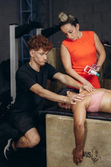
Initial care focuses on the RICER protocol – Rest, Ice, Compression, and Elevation – to minimize inflammation and pain following an injury.
Rehabilitation progresses through isometric exercises, dynamic stretches, and anti-inflammatory treatments for optimal healing.
RICER Protocol (Rest, Ice, Compression, Elevation)
The RICER protocol is the cornerstone of immediate sports injury management, crucial for minimizing damage and initiating the healing process. Rest involves immediately ceasing activity to prevent further strain on the injured anatomical structures – be it muscle, ligament, or bone.
Ice application, typically for 15-20 minute intervals, constricts blood vessels, reducing inflammation and pain. Compression, using an elastic bandage, helps control swelling and provides support. It’s vital to avoid overly tight wrapping.
Finally, Elevation, raising the injured area above heart level, aids in fluid drainage and further reduces swelling. Consistent application of RICER in the initial 24-72 hours is paramount for optimal recovery, setting the stage for subsequent rehabilitation phases. Understanding the anatomical basis of the injury informs how diligently each component of RICER is applied.
Immobilization Techniques
Immobilization plays a critical role in sports injury recovery, protecting damaged anatomical structures and facilitating healing. Techniques range from simple splints and slings to casts, depending on the injury’s severity and location. The goal is to limit movement at the injured joint or bone, preventing further aggravation.
For fractures, casts are often necessary to maintain alignment during bone remodeling. Ligament or tendon injuries may benefit from bracing or splinting, providing support while allowing controlled range of motion as healing progresses.
Proper immobilization considers the specific anatomy involved; incorrect application can hinder recovery or cause complications. Duration varies based on the injury type and individual healing rate, guided by medical professionals. It’s a temporary measure, transitioning to rehabilitation exercises once sufficient stability is regained.
Isometric Exercises for Early Rehab
Isometric exercises are foundational in early sports injury rehabilitation, focusing on muscle contraction without joint movement. This is crucial when motion is painful or contraindicated, preserving muscle strength and minimizing atrophy around the injured area. They activate muscle fibers, promoting blood flow and initiating the healing process.
For example, quadriceps sets after a knee injury, or gluteal squeezes for hip rehabilitation, are common starting points. These exercises target specific muscle groups, engaging them against an immovable resistance – often the patient’s own effort.
Isometric training helps maintain neuromuscular pathways, preparing the muscles for more dynamic movements later in rehab. They are generally pain-free and can be incorporated early, bridging the gap between acute injury and progressive loading.
Dynamic Stretches for Improved Flexibility
Dynamic stretches are controlled movements that take joints through their full range of motion, preparing muscles for activity and enhancing flexibility – a vital component of sports injury rehabilitation. Unlike static stretching, which holds a position, dynamic stretches mimic sport-specific actions, improving functional movement patterns.
Examples include leg swings, arm circles, and torso twists. These movements increase blood flow to muscles, elevating tissue temperature and reducing stiffness. They actively engage the neuromuscular system, improving coordination and proprioception – the body’s awareness of its position in space.
Incorporating dynamic stretching into a rehab program helps restore optimal muscle length and joint mobility, reducing the risk of re-injury and facilitating a return to sport. They are best performed after a warm-up and before more strenuous activity.
Anti-inflammatory Medication & Pain Management
Anti-inflammatory medication plays a crucial role in managing pain and reducing inflammation following a sports injury, often complementing other rehabilitation strategies. Nonsteroidal anti-inflammatory drugs (NSAIDs), both topical and oral, can effectively alleviate discomfort and swelling, allowing for greater participation in rehab exercises.
However, medication should be considered part of a broader approach, not a sole solution. It’s vital to address the underlying anatomical issue through targeted exercises and manual therapy. Pain management also involves techniques like ice, compression, and elevation (RICER protocol).
Understanding the potential side effects of NSAIDs and consulting with a healthcare professional is paramount. Effective pain control facilitates adherence to the rehabilitation program, promoting optimal healing and a faster return to activity.